-
动态


作者:Prof. Mayer
Abstract
摘 要
“The past is the mother of the future”
Henri Cartier Bresson, French Photographer,1908-2004
"过去是未来之母"
亨利·卡蒂尔-布雷松,法国摄影家,1908-2004
Introduction
简 介
Development and progress in spinal surgery have always been characterized by “back-and-forth movements” in clinical applications of technical innovations. Most evolutionary technical improvements which seemed to have a logical indication spectrum, with adequate feasibility and a perspective to improve early or late outcomes, have sooner or later become “standard” with a worldwide market penetration. A good example of such a development is anterior cervical discectomy and fusion (ACDF). It all started with the Cloward and Smith-Robinson technique , which was improved with the development of plates to support and fix the bone grafts. The bone grafts were replaced by cages made from different materials, and further technical improvement has led to the use of cages as stand-alone devices recently. This is a typical simple example of a continuous evolution of a surgical technique.
The lesson we can learn from this is that if a technical improvement follows the needs of the surgeon and if it improves or standardizes a surgical technique and its outcomes, the acceptance among the surgical community will be logical and high.
脊柱外科的发展和进步总是以技术创新在临床应用中的“来回运动”为特征的。大多数有变革性的手术技术发展中似乎都有一个合理的指征范围,具有可行性高且可改善早期和晚期效果的前景,迟早它们会成为渗透全球市场的“标准”。前路颈椎间盘切除融合术(ACDF)是诠释这种发展历程的一个好例子。这一切都始于Cloward和Smith-Robinson技术,随着支持和固定移植骨钢板的发展,该技术得到了改进。骨移植被不同材料制成的cage取代,技术的进一步改进可实现cage作为独立的设备使用。这是一个典型且简单例子阐释了外科技术将不断发展的特性。
我们从中可以学到的是,如果一项技术的发展是紧随外科医生的需求,且这发展改进或标准化一项技术及其结果,那么外科界的接受度将会很高。
History of Lumbar Disc Surgery
腰椎间盘手术的历史
Part 1: From Complete Laminectomy to Microsurgical/Microendoscopic Techniques
第一部分:从全椎板切除术到显微外科/显微内窥镜技术
The history of lumbar discectomy and lumbar decompression is one of the most fascinating chapters of spine surgery which has taught us a number of important lessons.
It was in 1909 when Krause and Oppenheim described the first lumbar discectomy (Figure 1). Erroneously they described the herniated disc as a chondroma of the lumbar spinal canal. Only 2 years later Goldthwaite and Middleton were the first to describe a herniated nucleus pulposus as a reason of low back pain and sciatica (Figure 2)
腰椎间盘切除术和腰椎减压术的历史是脊柱外科发展中最精彩的篇章之一,它给我们带来许多重要的启示。
1909年,Krause和Oppenheim首次报道了腰椎间盘切除术(图1)。他们错误地将椎间盘突出描述为腰椎管内的软骨瘤。仅仅2年后,Goldthwaite和Middleton首次将髓核突出描述为腰痛和坐骨神经痛的原因(图2)。
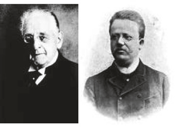
Figure 1: F Krause and H Oppenheim: first surgical removal of a “chondroma” of the spinal canal 1909.
图1:F Krause和H Oppenheim:1909年首次手术切除椎管内的“软骨瘤”。
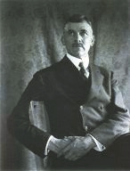
Figure 2: JE Goldthwaite: first description of herniated nucleus pulposus as reason for sciatica, 1911.
图2 :JE Goldthwaite:1911年首次描述髓核突出是坐骨神经痛的原因。
And it took another 11 years until Adson came up with the first report about surgical removal of herniated nucleus pulposus (Figure 3).
又过了11年,Adson提出了第一篇关于手术切除突出髓核的报告(图3)。
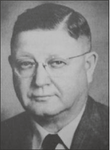
Figure 3: AW Adson: first description of surgical removal of herniated nucleus pulposus, 1922.
图3 AW Adson:1922年首次报道手术切除突出髓核。
However, like very often in medical history the merits for the first disc surgeries went to two other colleagues, namely, Mixter and Barr, who still are considered as having been the “first disc surgeons” in 1934 (Figure 4). They actually published the first series of successful disc operations in 1934. Their technique however was a complete laminectomy and some of the disc herniations were removed through a transdural approach.
当然,就像医学史上经常发生那样,第一例椎间盘手术的功劳归于另外两位医生,即Mixter和Barr,他们仍被认为是1934年进行“第一例椎间盘手术的外科医生”(图4)。实际上,他们在1934年报道了第一批成功的椎间盘手术系列,但他们是进行全椎板切除术,并且部分椎间盘突出是经硬膜入路切除的。
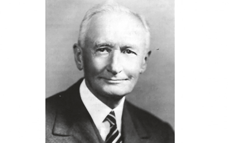
Figure 4: WJ Mixter: first case series of surgical removal of herniated discs 1934.
图4:WJ Mixter:1934年第一批椎间盘切除手术系列
It was obvious from the beginning that this was a very traumatic approach with the potential of a variety of complications including dural leaks and segmental instability as well as disabling back pain.
The search for less damaging approaches had started. Only 5 years later, Love described the first interlaminar approach which became the standard procedure for many years (Figure 5). But even though the rate of major surgical complications dropped over time, the problem of postoperative back pain and rapid progression of disc degeneration due to aggressive disc removal affected the clinical outcomes.
从一开始就很明显,这是一种非常具有创伤性的手术入路,有可能出现各种并发症,如硬脑膜渗漏、节段性不稳以及伤残性背痛。
人们开始寻找侵入性较小的手术入路。仅仅5年后,Love报道了首次经椎板间入路,这成为多年来的标准术式(图5)。但是,尽管主要手术并发症的发生率随着时间的推移而下降,但术后背痛和因激进的椎间盘切除而导致椎间盘退变迅速的问题影响了临床结果。
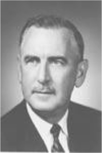
Figure 5: JG Love: first description of interlaminar approach, 1939.
图5:JG Love:1939年首次报道了经椎板间入路
While surgery led to a significant improvement of nerve root compression signs, patient satisfaction was impaired by symptoms which were due to the collateral damage the surgeon had produced. Interestingly this fear is still immanent in today's public opinion about disc surgery.
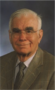
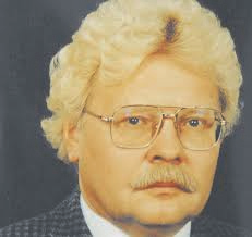
The reduction of collateral damage was the driving force for the two pioneers of lumbar microsurgery. In the same year 1977 Yasargil and Caspar described independently a microsurgical interlaminar approach , Figures 6(a) and 6(b). One year later, it was “Tex” Williams who was the first surgeon to perform this approach in the US. The pioneering work of JA McCulloch made this approach popular in the 90s of the last century and it has become a “gold standard” at least in the neurosurgical community worldwide. Other approaches such as the lateral extraforaminal access have been described in this book as well.
虽然手术明显的改善了神经根受压症状,但由于外科医生造成的附带损伤,病人的满意度受到了影响。有趣的是,这种恐惧在今天关于椎间盘手术的公众舆论中仍然存在。
减少附带损伤是两位腰椎显微外科先驱者的动力。同年1977年Yasargil和Caspar独立地描述了显微外科椎间板入路,图6(a)和6(b)。一年后,"Tex " Williams成为美国首次采用上述入路的外科医生。JA McCulloch的开创性工作使这种入路在上世纪90年代流行起来,至少在全球神经外科界,它已成为 "黄金标准"。他写的这本书还介绍了其他入路,如外侧椎间孔外入路。
(a)
(b)
Figure 6 : (a) G Yasargil, (b) W Caspar: first description of microsurgical interlaminar approach.
图6 (a) G Yasargil, (b) W Caspar: 首次描述显微外科椎板间入路。
“Microendoscopic discectomy” was described in the beginning of this century as a modification of the microsurgical technique where the surgical microscope is replaced by “open” endoscopy. This technique however did not add any further technical or clinical advantages. However both minimally invasive techniques are practiced with good and reproducible clinical outcomes.
本世纪初,“显微内窥镜椎间盘切除术”被描述为显微外科技术的改进,手术显微镜被“开放式”内窥镜取代。然而,这种技术并没有增加任何进一步的技术或临床优势。但是,这两种微创技术在实践中获得良好且可重复性的临床效果。
Lessons Learnt from Microsurgical Techniques
从显微外科技术中学到的经验教训
In summary lumbar microsurgery has significantly improved clinical short-term outcomes of lumbar discectomy mainly by reducing iatrogenic collateral damage. Thus, hospitalization times have become shorter, postop pain levels are lower, and intraoperative blood loss as well as the risk of infection is less.
Even though the advantages are obvious, several lessons had to be learnt by the protagonists of such techniques.
Since there is obviously no effect on the long-term outcome of lumbar discectomy, the acceptance especially by the older generation of spine surgeons has been low despite the obvious advantages.
It has been known for many years that long-term outcome of lumbar discectomy has different predictors than the short-term outcome. This is due to the fact that there is a progressive degeneration of the spine which can cause clinical symptoms at other levels which are not related to a previous disc surgery.
However we have learnt that one of the strongest predictors of a good long-term outcome is a good short-term outcome. And we have also learnt that a good short-term outcome is predicted by 2 factors: (1) the efficacy of nerve root compression and (2) the extent of iatrogenic collateral damage to muscles, ligaments, facet joints, nerve, and epidural space.
综上所述,腰椎显微手术主要通过减少医源性附带损伤而显著改善了腰椎间盘切除术的临床短期疗效。术后住院时间缩短,术后疼痛程度降低,术中出血量以及感染的风险也降低。
尽管这种技术的优势是显而易见的,但操作这些技术的主角们必须从中吸取一些经验教训。
由于腰椎间盘切除术的长期疗效没有明显改进,尽管有明显的优势,但脊柱外科医生,特别是老一代的,对此技术的接受程度很低。
众所周知,腰椎间盘切除术的长期疗效与短期疗效的预测因素不同。这是由于脊柱的进行性退变会导致其他水平的临床症状出现,而这些症状与之前的椎间盘切除手术无关。
但是,我们了解到,良好的短期疗效是预测长期疗效的最有力因素之一。我们还了解到,良好的短期疗效是取决于两个因素:(1)神经根压迫的效果;(2)医源性侧支对肌肉、韧带、关节突、神经和硬膜外间隙的损伤程度。
Part 2: The “Parallel World” of “Percutaneous” and Endoscopic Techniques
第二部分:“经皮”与内窥镜技术的“平行世界”
It was in 1964 when Lyman Smith published a paper about enzymatic dissolution of the nucleus pulposus, a procedure which he called chemonucleolysis. It was known at that time that an enzyme called Chymopapain, which was derived from the papaya plant, was able to hydrolyze proteoglycans. During experimental work in the 50s of the last century about the effects of papain, there was an interesting incidental finding. Intravenous injection of papain in rabbits resulted in a reversible collapse of rabbit ears, a finding which suggested an effect of this enzyme on cartilage. Similar effects were then reported on cartilage of joints, trachea, larynx, and bronchi. Since further studies on rabbits had shown that this enzyme dissolves the nucleus pulposus, it was Lyman Smith’s idea that an application in contained disc herniations could lead to an “intradiscal decompression”, thus relieving the symptoms from nerve compression due to a bulging lumbar disc.
In the 1980s this procedure became popular as the least invasive technique to treat herniated lumbar discs.
Mid- to long-term outcomes were good, complications were rare, and chemonucleolysis seemed to become a viable alternative to surgical discectomy.
Then something happened which was more a psychological phenomenon than rational based medical evolution. In the 70s, Hijikata, a Japanese surgeon, was fascinated by the posterolateral access to the disc space which was, at that time, in the pre-CT and pre-MRI era, very popular to perform diagnostic discographies (Figure 7). He developed tubes through which he could introduce this approach down to the posterolateral annulus under fluoroscopic control. With special trephines he could perforate the annulus and, using pituitary rongeurs, he could perform what he called “percutaneous nucleotomy”. He published this procedure in a regional scientific journal in Japanese language. This was one of the reasons why this procedure did not gain widespread attention among the surgical community but it was the birth of “percutaneous” and, later, endoscopic discectomy.
1964年,Lyman Smith发表了一篇关于酶溶解髓核的论文,他把这个过程称为化学核溶解。当时人们知道一种名为木瓜凝乳蛋白酶的酶(Chymopapain),这种酶是从木瓜植物中提取的,能够水解蛋白聚糖。在上世纪50年代关于木瓜蛋白酶作用的实验中,有一个有趣的意外发现。在兔子身上静脉注射木瓜蛋白酶会引起兔子耳朵的可逆性塌陷,这一发现表明这种酶对软骨是有影响。对关节、气管、喉和支气管的软骨也有类似的影响。由于对兔子的进一步研究表明这种酶可以溶解髓核,Lyman Smith的想法是,在椎间盘突出的地方应用这种酶可实现“椎间盘内减压”,从而缓解由于腰椎间盘突出引起的神经压迫症状。
在20世纪80年代,这种方法作为治疗腰椎间盘突出症中损伤最小的技术而流行起来。
中期、长期疗效良好且并发症少,化学髓核溶解术似乎成为一个可替代椎间盘切除术的选择。
随后发生的事情,更多的是一种外科医师对手术技能的执着,而不是基于理性的医学演变。在70年代,日本外科医生Hijikata对通过后外侧入路进入椎间盘间隙非常着迷,当时,在CT 和MRI之前的年代,这是非常流行的诊断椎间盘造影(图7)。他研发了一些管子,在透视下,他可以通过这种入路进入后外侧环。使用特殊的环钻在椎环上打孔,并用垂体咬骨钳,他可进行他所谓的“经皮髓核切除术”。他在一家地区性科学杂志上用日语发表了这一手术过程。这也是这种手术没有在外科界获得广泛关注的原因之一,但它是“经皮”以及后来的内窥镜椎间盘切除术诞生的开始。
Figure 7: Hijikata: first percutaneous nucleotomy, 1975.
图7 Hijikata:1975年第一次进行经皮髓核切除术。
It was the great merit of Parviz Kambin a Philadelphian spine surgeon to further develop this procedure in the 1980s (Figure 8).
Parviz Kambin是费城的一位脊柱外科医生,他在20世纪80年代进一步发展了这一手术操作,这是他的一大功绩(图8)。
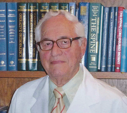
Figure 8: P Kambin: percutaneous discectomy, 1986.
图8 P Kambin:1986年进行经皮椎间盘切除术。
It is the “Kambin triangle” (the safe corridor to the lumbar disc between the exiting nerve root and the superior facet) which reminds us of his pioneering work (Figure 9).
这是“Kambin三角”(出口神经根和上小关节之间可直入腰椎间盘的安全走廊),这让我们想起他所做过的开创性工作(图9)。
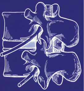
Figure 9: Kambin’s triangle for a safe posterolateral approach.
图9:安全后外侧入路的Kambin三角。
Schreiber, Suezawa, and Leu were the first to have the idea to perform this percutaneous nucleotomy under visual control using and endoscope (discoscopy) .
The author of this review adopted this technique, refined the instrument set (Figure 10), and published the results of a randomized controlled trial comparing microdiscectomy with endoscopic posterolateral discectomy.
Schreiber、Suezawa和Leu是最先有这个想法的人,他们提出在可视控制和内窥镜(椎间盘镜)下进行经皮髓核切除术。
这篇综述的作者采用了这一技术,改进了器械组(图10),并发表了一项比较显微椎间盘切除术与内窥镜后外侧入路椎间盘切除术的随机对照试验结果。
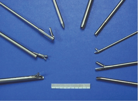
Figure 10: Early Instrument set for percutaneous endoscopic discectomy.
图10:经皮内窥镜椎间盘切除术的早期器械组。
A more lateral access route was described by Hal Mathews and Tony Yeung in the second half of the 1990s.
This lateral extraforaminal approach enabled the removal of far lateral disc herniations as well as more medially located pathologies because the approach corridor was more parallel to the posterior rim of the annulus (Figure 11).
20世纪90年代后半期,Hal Mathews和Tony Yeung描述了一种更横向的通道。
外侧椎间孔外入路可以切除远外侧的椎间盘突出以及更内侧的病变,因为此入路通道更平行于椎间盘环的后缘(图11)。
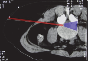
Figure11: Approach corridor and visual field for transforaminal approach.
图11:经椎间孔入路的通道和可视范围。
Lessons Learnt
经验学习
The indication spectrum for posterolateral and transforaminal endoscopic techniques was limited, which was one of the reasons why endoscopic discectomy remained at a low level of acceptance among spine surgeons in the 1980s and 1990s.
There were other reasons: the variety of instruments was limited, the optical systems were not as good as nowadays, and the technical advantages as compared to microsurgery were small.
后外侧和经椎间孔内窥镜技术的指征范围是有限的,这是80年代和90年代内窥镜下椎间盘切除术在脊柱外科医生中接受程度低的原因之一。
其他原因还有:手术器械种类有限,光学系统不如现在好,与显微外科手术相比,技术优势不大。
Part 3: From a Nondisruptive to a Disruptive Surgical Technology
第三部分:从非颠覆性到颠覆性的外科技术
But what was the missing link or major step? The answer is simple: endoscopy was used in a “dry” environment because the technical advantages of joint arthroscopy were not applied.
Whereas in joint arthroscopy surgical dissection was performed “under water” with continuous irrigation and suction, this principle was not applied in the spine because of the erroneous assumption that irrigation might not be of help or necessary in non-preformed anatomic spaces. The advantages of continuous irrigation (hemostasis, flushing of small bleeding, identification of the bleeding source, better identification of microanatomy, and separation of tissue layers by simple irrigation) were not realized.
Moreover, the technique focused on lateral extraforaminal approaches, and the most traditional interlaminar approach was believed not to be feasible with such a technique.
This is why “the first wave” of lumbar endoscopic techniques remained a nondisruptive technology.
Things changed in the late 90s. It was the merit of Anthony Yeung who started to consequently apply arthroscopic technology for transforaminal as well as interlaminar approaches (Figure 12).
但这当中是缺少了什么环节或主要步骤?答案很简单:因为关节镜的技术优势没有被应用,内窥镜是在一个“干燥”的环境中使用的。
关节镜手术是在“水下”通过持续冲洗和抽吸进行的,而这一原理并没有应用于脊柱手术中,因为人们错误地认为冲洗在非预制的解剖空间里可能没有帮助也没必要。连续冲洗的优点(止血、冲洗少量出血、识别出血源、更好地识别微观解剖结构、通过简单冲洗分离组织层)均无实现。
此外,该技术倾向于外侧椎间孔外入路,而最传统的经椎板间入路被认为在内窥镜下是不可操作的。
这就是为什么腰椎内窥镜技术的“第一波浪潮”仍然是一种非颠覆性的技术。
在90年代末,事情开始发生转变。这是Anthony Yeung的功劳,他逐渐将关节镜技术应用于经椎间孔以及经椎间板入路(图12)。

Figure 12: A Yeung: first application of transforaminal approach under continuous irrigation.
图12:A Yeung:持续灌洗下经椎间孔入路的首次应用
There were three major steps, which transferred spinal endoscopy into a disruptive technology:
(1) “under-water-dissection”: continuous irrigation reduced intra- and postop bleeding and infection rates and significantly improved visibility of anatomic structures;
(2) the range of approaches increased from pure transforaminal or posterolateral to interlaminar because
(3) rongeurs, high-speed drills, and other instruments could be used.
Success rates increased and recurrence rates decreased. Rapidly this technology was adopted mainly in Asian countries.
At the beginning of the 2000s it was Sebastian Rütten, a German spine surgeon, who adopted this technology and applied it for interlaminar endoscopic approaches. This significantly enlarged the indication spectrum of this technology (Figure 13).
将脊柱内镜技术转变为一项颠覆性技术的主要步骤有三个:
(1)“水下解剖”:持续冲洗减少了术中和术后的出血及感染率,并大大提高了解剖结构的可见度;
(2)手术入路范围从单纯的经椎间孔或后外侧扩大至经椎间板;因为
(3) 可以使用骨钳、高速磨钻和其他器械。
成功率提高,复发率降低。这项技术很快被采用,特别是在亚洲国家。
在21世纪初,德国脊柱外科医生Sebastian Rütten采用了这项技术,并将其应用于经椎板间内窥镜手术。这极大地扩大了这项技术的适应症范围(图13)。

Figure 13: S Rütten: first interlaminar approach and application of arthroscopic technique.
图13 S Rütten:首次经椎板间入路和应用关节镜技术
The current indication spectrum for thoracic and lumbar applications is wide and covers all types of degenerative (and other) pathologies which have been a domain of microsurgical techniques in the past (Table 1)
目前胸腰椎应用的适应症范围很广,涵盖了所有类型的退行性(和其他)病理(表1),在过去这些是属于显微外科技术领域的。

Table 1
Indications for full-endoscopic posterior/lateral thoracic and lumbar spine surgery.
(i) Decompression of central and foraminal spinal stenosis
(ii) Decompression of lateral recess stenosis
(iii) Removal of all types of disc herniations incl. difficult cases and recurrent disc herniations
(a) Medial disc herniations
(b) Down migrated disc herniations
(c) Bilateral disc herniations
(d) Recurrent disc herniations
(e) Calcified disc herniations
(iv) Removal of synovial cysts
(v) Removal of epidural hematoma
(vi) Removal of thoracic disc herniations and decompression of thoracic stenosis
(vii) Palliative decompression metastases
表1
全内窥镜下胸腰椎后/侧入路手术的适应症。
(i) 中央和椎间孔椎管狭窄减压
(ii) 侧隐窝狭窄减压
(iii) 切除所有类型的椎间盘突出,包括复杂病例和复发性椎间盘突出症
(a) 内侧椎间盘突出症
(b) 下移的椎间盘突出症
(c) 双侧椎间盘突出症
(d) 复发性椎间盘突出症
(e) 钙化椎间盘突出症
(iv) 切除滑膜囊肿
(v) 清除硬膜外血肿
(vi) 胸椎间盘突出症摘除和胸椎管狭窄症减压
(vii) 转移瘤的姑息性减压
Summary
总 结
The first attempts of endoscopic lumbar spine surgery date back to the early 1980s. However, only in the last decade this technology has become a disruptive technology with the potential to replace microsurgical techniques especially for degenerative lumbar spine disorders.
The strong input and high acceptance among Asian spine surgeons have triggered a very dynamic clinical and scientific workflow on this topic. A PubMed search for scientific publications on endoscopic lumbar spine surgery shows that more than 80% of the publications have their origin in Asian countries. It has been shown that even though there is a certain learning curve for endoscopic techniques, once the surgeon is familiar with it, he can achieve comparable and sometimes better clinical results as conventional microsurgical operations.
The complication rates of experienced and well-trained surgeons are low.
The iatrogenic collateral damage of the different approaches to the lumbar spine is diminished and most of the procedures can be performed in an outpatient setting.
腰椎内窥镜手术的首次尝试可以追溯到20世纪80年代初。但直到最近十年,这项技术才成为一项颠覆性的技术,才有可能取代显微外科技术,特别是对退行性腰椎疾病。
亚洲脊柱外科医生的激情投入和高度接受,引发了关于这一主题的非常活跃的临床和科学工作潮。在PubMed上搜索有关腰椎内窥镜手术的科学出版物,发现80%以上的出版物都来自于亚洲国家。事实证明,尽管内窥镜技术有一定的学习曲线,但一旦外科医生熟悉了它,他就可以取得与传统显微外科手术相当的临床效果,有时甚至更好。
经验丰富、训练有素的外科医生手术的并发症率很低。
不同入路下进行的腰椎手术医源性附带损伤减少,且大多数手术可以在门诊环境进行。
The Future
未 来
Today we are in a stage which I would call “microendoscopic blending” where the dynamics of technical improvement of endoscopic techniques suggests that the overlap of indications for this technology vs. microsurgery will step by step convert into a scenario where endoscopic techniques replace microsurgical techniques. The great challenge is the learning curve and the training of young surgeons. The acceptance of this technology is high among young surgeons but it is the task and duty of the protagonists of the older generation, the hospitals, and the scientific societies to develop learning- and training-concepts to shorten learning curves and to improve technical quality and clinical outcomes.
今天,我们正处于一个我称之为“显微内窥镜融合”的阶段,内窥镜技术的发展动态表明,这种技术与显微外科手术的适应症重叠将逐步转变为内窥镜技术取代显微外科技术的局面。最大的挑战是学习曲线和对年轻外科医生的培训。年轻的外科医生对这项技术的接受程度很高,所以老一代的外科医生、医院和科学学会的任务和职责是发展学习和培训概念,以缩短学习曲线,提高技术质量和临床疗效。
